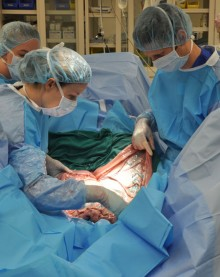
CALEC surgery represents a groundbreaking advancement in the treatment of corneal damage, offering renewed hope for patients facing vision loss from untreated eye conditions. Developed at the prestigious Mass Eye and Ear, this innovative procedure utilizes cultivated autologous limbal epithelial cells, or CALEC, to restore the cornea’s surface with remarkable efficacy. By extracting stem cells from a healthy eye and cultivating them into a graft, CALEC surgery has achieved over 90 percent success rates in restoring corneal surfaces in clinical trial participants. This pioneering stem cell therapy not only facilitates corneal surface restoration but also addresses the underlying issues of limbal stem cell deficiency in patients suffering from various types of ocular trauma. As research continues to unfold, CALEC surgery may signal a new era in corneal damage treatment, making previously untreatable conditions manageable and improving the quality of life for countless individuals suffering from severe eye injuries.
Known as cultivated limbal epithelial cell transplantation, CALEC surgery is a revolutionary technique that reinvigorates the healing potential of the eye. By harnessing the power of stem cells, specifically limbal epithelial cells sourced from a healthy donor eye, this method aims to rejuvenate the corneal surface effectively. This approach not only enhances corneal restoration but also serves as a vital solution for those affected by severe ocular injuries. The implications of this treatment extend beyond mere visual rehabilitation, providing patients with relief from chronic pain and restoring their ability to engage fully with their surroundings. As attention grows around corneal surface restoration techniques, CALEC emerges as a beacon of hope in the field of ophthalmology.
The Breakthrough of CALEC Surgery
The recent introduction of cultivated autologous limbal epithelial cells (CALEC) surgery marks a significant milestone in the field of ophthalmology. This innovative procedure, developed at Mass Eye and Ear, leverages stem cell therapy to restore the corneal surface, providing renewed hope for patients suffering from severe corneal damage. Utilizing limbal epithelial cells harvested from a healthy eye allows surgeons to regenerate the damaged areas, drastically improving patients’ quality of life and restoring their vision, which was previously thought irreparable. This groundbreaking surgery is a testament to the potential of stem cell therapies in treating complex eye conditions.
Ula Jurkunas, a leading figure in this pioneering work, has indicated that CALEC surgery has demonstrated an impressive success rate exceeding 90% in restoring the corneal surface after transplantation. Over an 18-month monitoring period involving 14 patients, the therapy not only proved to be safe but also exhibited remarkable efficacy, signifying a leap forward in the treatment landscape for corneal injuries. As more comprehensive trials are planned, the hope is to solidify CALEC as a standard treatment option for patients facing corneal challenges.
Understanding Corneal Damage and Its Treatment
Corneal damage can arise from various causes, including chemical burns, infections, or traumatic injuries, leading to what is known as limbal stem cell deficiency. In such cases, the cornea fails to regenerate due to the depletion of essential limbal epithelial cells responsible for maintaining its smooth surface. This condition often leaves patients with chronic pain and visual impairments, making traditional corneal transplants unfeasible. The successful application of stem cell therapy through CALEC surgery offers an innovative solution to address these severe eye injuries by rejuvenating the corneal surface.
The treatment involves precisely extracting limbal epithelial cells from a donor eye, cultivating them in a lab, and then transplanting them into the affected eye, thus restoring its functionality. This cutting-edge approach not only highlights the capabilities of stem cell therapy for eyes but also signifies a paradigm shift in how medical professionals can manage corneal damage. As research continues, the potential for developing more advanced and widely applicable treatments grows, opening new avenues for those who previously had limited options.
The Role of Stem Cell Therapy in Ophthalmology
Stem cell therapy represents a transformative approach in treating various eye disorders, particularly in the restoration of corneal surfaces. The technique’s foundation lies in the ability to harness the regenerative properties of limbal epithelial cells, which play a crucial role in maintaining corneal integrity. By employing this technology, researchers and clinicians can offer a solution for patients afflicted with conditions that leave them with inoperable corneal surfaces, which traditional methods cannot address effectively. This innovative approach through CALEC surgery bridges the gap in current treatment methodologies.
Recent clinical trials have underscored the potential of stem cell therapies, particularly those implemented at institutions like Mass Eye and Ear. As researchers explore further enhancements and broader applications, the integration of stem cell-based treatments such as CALEC is poised to redefine ocular treatment paradigms. The ongoing development and refinement of these therapies promise to expand treatment options for various ocular conditions, ultimately improving outcomes for patients with corneal damage.
Innovative Approaches to Eye Care
Innovative approaches in eye care are crucial for redefining treatment methodologies, particularly for conditions that have historically been deemed untreatable. The CALEC procedure exemplifies the convergence of modern science and clinical application, showcasing how advanced techniques can lead to successful patient outcomes. With the backing of extensive research and trials, the progress made in cultivated limbal epithelial cell surgery is encouraging, as it opens the door to vital advancements in treating corneal injuries.
As the medical community continues to address the challenges presented by corneal damage, the promise of stem cell therapy becomes increasingly evident. Future studies and clinical trials, especially those focusing on larger sample sizes and controlled environments, will be vital for cementing CALEC surgery as a widely accepted treatment modality. This innovative spirit within eye care not only fosters hope among patients but also propels the medical field toward more comprehensive and effective interventions in ocular health.
Future Directions of CALEC Studies
With the successful initiation of CALEC surgery trials at Mass Eye and Ear, the future directions for this innovative treatment look promising. Researchers are advocating for larger-scale studies that involve diverse patient populations to gather more comprehensive data regarding the effectiveness and safety of CALEC surgery. Such studies are essential for understanding the long-term implications of using stem cell-based therapies in ocular applications, and they will play a crucial role in pushing for FDA approval, making these treatments accessible to those in need.
Moreover, the development of an allogeneic manufacturing process for CALEC could significantly broaden the potential for treatment, as it would present opportunities to assist patients with bilateral corneal damage. This advancement would not only enhance the feasibility of the procedure but also reflect a commitment to pushing the boundaries of ocular regenerative therapies. As researchers continue to collaborate across institutions, the synthesis of innovative techniques and robust clinical trials is expected to pave the way for enhanced outcomes in corneal damage treatment.
The Safety Profile of CALEC Surgery
A critical aspect of any new medical procedure is its safety profile, especially with novel treatments like CALEC surgery. The clinical trials conducted so far have demonstrated a favorable safety record, with no serious adverse effects reported among participants. These findings underscore the importance of thorough preclinical testing and careful patient monitoring during and after the transplantation process. The low incidence of complications highlights the commitment of researchers and clinicians to ensuring patient safety as a priority throughout the trial phases.
Despite its promising safety outcomes, the procedure is still classified as experimental, and further studies are essential to assess long-term effects of CALEC therapy. Understanding the nuances of potential risks associated with stem cell grafts, particularly concerning infections or other complications, is vital for developing robust treatment protocols. Continued research in this area will not only reinforce the safety of this innovative approach but also instill confidence among patients seeking novel therapeutic options for corneal damage.
The Impact of CALEC on Visual Acuity
The primary goal of any ocular treatment, particularly for those suffering from corneal injuries, is to restore visual acuity. The clinical trial of CALEC surgery revealed notable improvements in vision among participants, with varying degrees of success reported within the study framework. As patients were monitored over an extended period, the researchers observed enhanced visual acuity alongside restored corneal integrity, solidifying the link between successful stem cell application and improved sight for individuals facing debilitating eye conditions.
Such improvements in visual acuity not only transform patients’ day-to-day lives but also contribute to the larger narrative of how innovative treatments can enhance overall quality of life. As eyecare professionals refine their approaches, the continued development of effective therapies based on stem cell technology may soon lead to breakthroughs that allow for even greater restoration of sight. The encouraging outcomes associated with CALEC surgery could inspire significant advancements in treating one of the most common and life-altering afflictions—corneal damage.
Expanding Access to CALEC Treatments
One of the central challenges in the rollout of any new medical treatment is ensuring access for all patients who may benefit from it. The advancements made with CALEC surgery at Mass Eye and Ear set a new benchmark in ocular care, yet it is crucial to develop a strategic plan for widespread accessibility. Aligning resources, funding, and infrastructure will be imperative to enabling patients nationwide to take advantage of this groundbreaking therapy.
Future initiatives may involve partnerships with healthcare systems across the country to facilitate training, establish treatment protocols, and expand the availability of CALEC surgery. By fostering collaboration among research institutes, hospitals, and regulatory bodies, the aim will be to create a harmonious environment that can quickly adapt to new interventions. This focus on access mirrors the broader goals of the medical community to provide equitable healthcare resources, ultimately benefiting all those affected by corneal damage.
The Role of Collaborative Research in CALEC Development
Collaboration is a pivotal factor in the success of innovative medical treatments, and the development of CALEC surgery exemplifies this principle in action. The interplay between researchers at Mass Eye and Ear and institutions such as Dana-Farber and Boston Children’s Hospital has led to necessary advancements in stem cell therapy for ocular applications. This collaborative spirit ensures that diverse expertise is leveraged, fostering an environment conducive to breakthroughs in patient care.
Moving forward, such partnerships will continue to be essential in addressing the complexities of corneal damage and refining treatment protocols. By committing to sharing knowledge and resources among various research teams, the ophthalmic community can expedite the advancement of therapies like CALEC. This collaborative approach marks a significant step toward creating comprehensive, multi-faceted solutions in the fight against vision impairment, further emphasizing the importance of unity in medical research.
Frequently Asked Questions
What is CALEC surgery and how does it help with corneal damage treatment?
CALEC surgery, or cultivated autologous limbal epithelial cells surgery, is a pioneering treatment developed at Mass Eye and Ear that involves harvesting limbal epithelial cells from a healthy eye, expanding them into a graft over a few weeks, and transplanting this graft into a damaged cornea. This ground-breaking procedure aims to restore the cornea’s surface, offering new hope for patients with corneal damage that has been deemed untreatable.
How effective is CALEC surgery for patients with corneal damage?
In clinical trials at Mass Eye and Ear, CALEC surgery demonstrated over 90% effectiveness in restoring the corneal surface. Specifically, the success rate for complete restoration reached 79% at the 12-month mark and was maintained at 77% at the 18-month follow-up, making it a promising option for those suffering from severe corneal damage.
What types of injuries can CALEC surgery effectively treat?
CALEC surgery is designed to treat corneal damage resulting from various injuries, including chemical burns, infections, and other trauma that leads to a depletion of limbal epithelial cells. This deficiency can cause chronic pain and vision challenges, which CALEC surgery aims to alleviate by restoring the corneal surface.
What are limbal epithelial cells and their role in corneal health?
Limbal epithelial cells are healthy stem cells located at the outer border of the cornea, known as the limbus. They are essential for maintaining the smooth surface of the eye. When these cells are depleted due to injury, it leads to limbal stem cell deficiency, resulting in permanent corneal damage that CALEC surgery seeks to rectify.
Is CALEC surgery currently available for all patients with corneal issues?
As of now, CALEC surgery remains experimental and is not widely available, including at Mass Eye and Ear. The procedure is under further study to gather more data for potential federal approval before it can be offered to a broader patient population.
What steps are involved in the CALEC surgical procedure at Mass Eye and Ear?
The CALEC surgical procedure involves multiple steps: first, a biopsy is taken from the healthy eye to extract limbal epithelial cells. These cells are then cultured in a lab to create a cellular graft over a period of 2-3 weeks. Finally, this graft is surgically transplanted into the damaged eye, aiming to restore its corneal surface.
What can patients expect in terms of recovery after CALEC surgery?
Patients undergoing CALEC surgery can expect variable recovery outcomes, with some experiencing significant improvements in visual acuity over 12 to 18 months. While most participants in the clinical trial reported benefits, each individual’s recovery may differ based on the extent of corneal damage and other factors.
What future advancements are anticipated for CALEC surgery?
Future advancements for CALEC surgery may include developing an allogeneic manufacturing process to use limbal stem cells from healthy donor eyes, potentially expanding treatment availability to patients with damage in both eyes. Ongoing research aims to include larger patient groups and longer follow-up periods to better assess the treatment’s efficacy and safety.
Are there any risks associated with CALEC surgery?
While CALEC surgery has demonstrated a high safety profile, as with any surgical procedure, there are risks involved. In the clinical trials, most adverse events were minor and resolved quickly; however, some patients experienced complications like bacterial infections linked to prolonged contact lens use.
How does CALEC surgery compare to traditional corneal transplants for vision rehabilitation?
Unlike traditional corneal transplants, which are standard care for vision rehabilitation, CALEC surgery specifically targets patients with limbal stem cell deficiency and aims to restore the corneal surface without the need for donor corneal tissue. This makes it a novel approach for patients who cannot receive standard corneal transplants.
| Key Points | Details |
|---|---|
| First CALEC surgery performed | Ula Jurkunas performed the first CALEC surgery at Mass Eye and Ear. |
| Clinical trial success | 14 patients successfully restored corneal surfaces with 90% effectiveness. |
| Procedure method | Involves extracting stem cells from a healthy eye, expanding them, and transplanting them into a damaged eye. |
| Effectiveness over time | 50% of participants had complete restoration at 3 months; increased to 79% and 77% at 12 and 18 month follow-ups, respectively. |
| Safety profile | No serious adverse events; one participant had a bacterial infection unrelated to the procedure. |
| Future goals | Aim to establish allogeneic manufacturing of limbal stem cells to broaden treatment availability. |
| Funding and collaboration | Study funded by National Eye Institute; collaboration with Dana-Farber and Boston Children’s Hospital. |
Summary
CALEC surgery represents a groundbreaking advancement in the field of ophthalmology, specifically in the treatment of corneal damage previously deemed untreatable. This innovative procedure, which utilizes stem cells to effectively restore the corneal surface, has shown promising results in clinical trials, significantly improving patient outcomes. With a high success rate and a strong safety profile, CALEC surgery not only offers hope to patients suffering from severe corneal injuries but also paves the way for further research and potential FDA approval, highlighting its pivotal role in future eye care treatments.
